
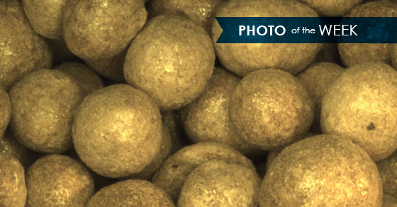
This week’s photo shows magnified pellets from the FEECO Innovation Center. The image was captured via a microscope, and stored through a high-tech software program.
FEECO’s lab technicians took the photo to see a more detailed look at the product they pelletized. Evaluation of a magnified, pelletized material can reveal much more than the naked eye. Color discrepancy is more detectable, which could indicate an uneven mixture of binder and material, or pellet surface may not be as smooth as desired for the end product. The microscope’s images help lab technicians to further fine-tune the agglomeration process used, in order to produce the desired results.
The microscope proves to be useful in many other areas as well. From comparative studies, to material feed analysis, the microscope’s magnified images are beneficial in each stage of the lab testing process.
The microscope works by taking shots at different focal points- top, middle, and bottom of the sample- and combining them to form one focused, magnified image. The photos provide concrete confirmation of the sample’s exterior and composition, revealing features often unrecognizable without magnification.
For more information on the microscope’s images, or FEECO’s capabilities in the Innovation Center, contact us today!